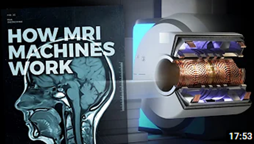
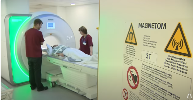
MRIs have completely changed how we view and understand our bodies. It has never been easier to visualize organs with such high details. We can safely locate and identify tumors in the kidneys, brain, stomach, and pancreas. We can inject paramagnetic contrast agents into the bloodstream to locate blockages in the heart, allowing doctors to accurately implant life-saving stents to open blood vessels. A tiny keyhole surgery that patients can recover from quickly, where once dangerous open-heart surgery was the only option. Magnetic resonance imaging has truly changed the nature of medical diagnosis and treatment. A technology that seems straight out of a sci-fi novel, how they work is a complete mystery to most.
To unlock the power of this machine physicists and engineers first had to discover and master the principles of quantum mechanics, superconducting magnets, computer science, and mathematics. The clean futuristic white facade of an MRI machine hides a world of complicated and marvelous engineering that you may not have ever considered. Check out this great video from the Real Engineering channel of YouTube from Keysight Technologies, a provider of electronic design and test solutions, to see for yourself. Here are a few excerpts:
“Prior to their introduction to medicine, we peered into our bodies using harmful ionizing x-rays or low-detailed ultrasounds. While incredibly useful, even to this day, these two imaging techniques pale in performance to the safe, millimeter resolution of MRIs. Capable of creating a 3D reconstruction of the body rather than a flat 2D image. Achieving all of that without any moving parts. Imaging in medicine relies on collecting signals from the body based on the innate physical properties of tissues. Ultrasound imaging relies on how sound waves bounce off tissues of different densities. X-rays form images based on the absorption of high-energy radiofrequency waves. Magnetic resonance imaging, however, relies on something far less intuitive, the quantum properties of the hydrogen atom. The human body is teeming with hydrogen atoms located in water, carbohydrates, and proteins. To image the body, MRI machines exploit a quantum property of these hydrogen atoms. A property called spin. How it does this is a marvel of modern physics and engineering.”
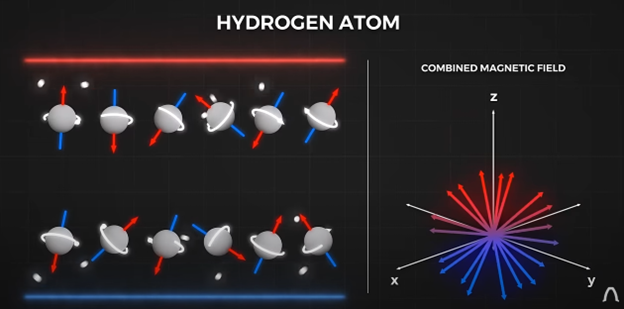
“Spin is an innate property of particles just like mass and charge. Spin makes particles behave like tiny bar magnets. The proton inside the hydrogen nucleus behaves like a magnet. The orientation of its magnetic north is described probabilistically. Under normal circumstances, this probability is evenly distributed. This means that the combined magnetic field of many hydrogen atoms cancels out. But, this changes when the hydrogens are inserted into a large external magnetic field just like the ones MRIs create.
This changes the distribution of the tiny magnets, augmenting the number of atoms aligned with the external magnetic field. This imbalance is the source of the MRI signal, as we can manipulate these tiny magnets to produce signals that can be processed into images.”
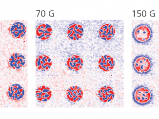
“Common MRI field strengths are 1.5 to 3 tesla, around 300,000 times stronger than the earth’s magnetic field and 30,000 times stronger than your common fridge magnet. You absolutely do not want anything ferrous around these machines during operation, a field this strong can lift nearby wheelchairs straight off the ground. For research purposes, MRIS can produce even higher magnetic fields, up to 20 teslas. Achieving this intense magnetic field comes as no easy feat. Early MRIs used permanent magnets as their source of the main magnetic field but only reached strengths of 0.5T limiting the resolution of the machine. Electromagnets can be used to reach stronger magnetic fields, but standard electromagnets can’t produce a 1.5 tesla field. Higher magnetic fields require higher electric currents that would melt ordinary wires. To achieve larger currents in the wires, engineers required superconducting coils.”
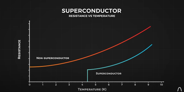
“Superconductors are another sci-fi technology. Temperature affects all metallic conductors. With resistance gradually lowering with temperature. But superconducting materials are special, in that their resistance drops to zero at temperatures close to minus 273 degrees Celsius, or absolute zero. In theory, when this happens, an electric current could travel in a loop of superconducting material indefinitely, never needing a power source.”
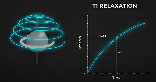
“We need a way of identifying different kinds of tissues to form an image, to do that we need to contrast the tissues using two different signal types. The first one deals with how quickly atoms re-align themselves with the large magnetic field after a nudging pulse. This is called T1 relaxation. The second measure comes from the physical reality of interaction between hydrogens.”

“Just like a photographer can play with camera settings to take pictures of either the bright sky or a dim environment, MRI technicians can also play with two settings to take images of contrasting tissues, the
time between pulse repetition, and how long to wait to listen for a signal. These two parameters are chosen by the technician each time an MRI is performed and they choose them depending on what the doctor would like to image. For example, T1 is used to image fatty tissues while suppressing the signal from water. But maybe the doctor wants to assess the cerebral spinal fluid in the spine or the brain, and for that T2 signals are emphasized to enhance the signal from water-based fluids. Remember, the signal is collected as a sum from a singular slice, The machine needs to form a 2D image of each slice. There is one extra layer of complexity since the receiver coil can only measure the sum of the signals from all the decaying hydrogens in the slice.”
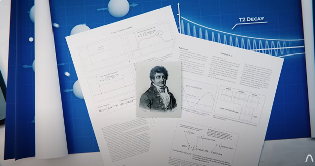
“This is yet another story where old mathematics discoveries guide modern technology. In 1822 Joseph Fourier created a mathematical framework that deconstructs complex waves into simple additions of simpler waves. This framework can be extended to 2D grayscale geometries. Just like a musical melody can be simplified into additions of simpler notes. Any image can be deconstructed into a weighted average of simpler black and white stripes. This is what MRIS uses to create images. Instead of sampling individual pixels, MRIs sample different striped patterns.”
For more info about Keysight, see www.keysight.com.
For more videos from Real Engineering, see Real Engineering – YouTube